


Contents
In line with its mission, the Editorial Board of MedTvoiLokony makes every effort to provide reliable medical content supported by the latest scientific knowledge. The additional flag “Checked Content” indicates that the article has been reviewed by or written directly by a physician. This two-step verification: a medical journalist and a doctor allows us to provide the highest quality content in line with current medical knowledge.
Our commitment in this area has been appreciated, among others, by by the Association of Journalists for Health, which awarded the Editorial Board of MedTvoiLokony with the honorary title of the Great Educator.
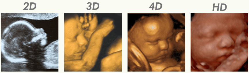
In conventional (2D) ultrasound, the image on the camera monitor is two-dimensional. For each of the structures depicted in this way, the transverse and longitudinal dimensions can be specified. On their basis, the computer calculates the volume of the organ. What is fetal imaging with 3D and 4D ultrasound? When is it worth doing?
What is the difference between traditional ultrasound and 3D and 4D ultrasound?
Technique images 2D ultrasound are sections of a given structure. This is not the case 3D techniques. It allows you to obtain data on three dimensions: length, width and depth of an organ. By composing this data, the computer recreates the three-dimensional appearance of the measured structure. The obtained images are still, presented frame by frame.
4D ultrasound it is used to dynamically present the image obtained with the 3D technique. As a result, the pregnant woman sees a three-dimensional image of the fetus moving inside her womb on the monitor screen.
Of course, 3D and 4D ultrasound examinations are very attractive for mothers who want to see their unborn babies. However, it should not be forgotten that apart from the possibility of obtaining souvenir photos for a child’s album, this diagnostics involves measurable medical benefits. First, the test record can be played back and analyzed multiple times. In addition, it allows for a precise assessment of the location and surroundings of individual anatomical structures. It also allows the use of invasive assisted reproductive techniques under ultrasound control.
When is 3D / 4D ultrasound recommended?
In the case of multiple pregnancies, 3D / 4D ultrasound is recommended as it is much more accurate than conventional 2D examination. It allows you to see all the fetuses, their relationship, location, number of placentas and amniotes.
Finally – both in multiple and single pregnancy – 3D / 4D ultrasound provides invaluable services detecting fetal defects. Once made, measurements allow for an infinite number of sections and projections, and for the re-analysis of images offline.
Undoubtedly, in the hands of an experienced doctor, 3D / 4D ultrasound machines provide invaluable services in the field of obstetric diagnostics. In the absence of contraindications to their implementation, they should therefore be recommended to every pregnant woman who can afford to bear their cost – at least once, between 18 and 22 weeks of pregnancy.
What will we see on 3D and 4D ultrasound?
Contrary to the traditional ultrasound performed during pregnancy, the 3D and 4D USG technology allows for obtaining an image that is clear for parents. Conventional ultrasound is a two-dimensional grayscale image that is understandable to the physician but may be unclear to the layman. Modern ultrasound technologies make it possible to obtain a realistic image of the baby in the womb.
By observing the fetus in three dimensions, we can see the clear shape of the baby, the surface of the body and even the shape of the face. There are anatomical details that are also helpful for the doctor who can more accurately assess how the baby is developing. However, the image obtained during the examination depends on several factors, such as the position of the fetus, the amount of amniotic fluid, the amount of body fat, and the gestational age.
In the case of 4D ultrasound, the image of the fetus is mobile, which means that we will see the baby’s movements, its behavior, and also facial expressions. A special type of ultrasound is the use of HD Live technology. It allows you to obtain a static or dynamic three-dimensional image that is more realistic. It is influenced by the ability to determine the direction of the virtual light falling on the child’s figure and face.
Also read about the following studies:
- Epidural ultrasound
- Gynecological ultrasound
- Pregnancy ultrasound
Waveform of 3D and 4D ultrasound
Performing a multidimensional ultrasound examination is similar to traditional ultrasound in pregnancy through the abdominal wall. It does not require any special preparation. It is enough to expose the abdomen, and the doctor will use a special gel to then apply the ultrasound machine head to the skin. We will then see a realistic picture of the child on the screen, and the gynecologist will assess his health. Duration of 3D and 4D ultrasound that’s ok 20-30 minutes.
After the visit, the patient can receive the obtained photos or record them on a DVD or transfer them to an external medium. Price for 3D or 4D ultrasound amounts to an average of PLN 200-300.
3D and 4D ultrasound in the assessment of birth defects
Based on 3D and 4D USG examinations, it is possible to more accurately diagnose abnormalities in the development of the fetus, diagnosed based on anatomical changes. Moreover, the multidimensional ultrasound allows the assessment of the baby’s organs as well as the blood flow. Congenital defects that can be detected by this test include:
- wrong body proportions;
- limb deformities, extra fingers;
- spine problems such as spina bifida;
- cleft lip;
- cleft palate;
- unusual face shape – especially the nasal bone can be an indicator of Down syndrome;
- neck translucency – this parameter may indicate many syndromes caused by chromosomal aberrations, including Down’s, Turner, Edwards and Patau’s syndromes, but it should be measured between 11 and 14 weeks of pregnancy;
- heart defects;
- defects of the kidneys and bladder;
- defects of the central nervous system.
The content of the medTvoiLokony website is intended to improve, not replace, the contact between the Website User and their doctor. The website is intended for informational and educational purposes only. Before following the specialist knowledge, in particular medical advice, contained on our Website, you must consult a doctor. The Administrator does not bear any consequences resulting from the use of information contained on the Website.