


Contents
4 weeks pregnant: what happens from conception, ultrasound, brown discharge
In the 4th week of pregnancy, the main symptom is a delay in menstruation. Subfebrile temperature is steadily increasing. The uterus is just beginning to grow. Now its size is the same as a hen’s egg. There are no pronounced symptoms of fertilization yet.
Changes in the 4th week of pregnancy
Pregnancy can be diagnosed at this time. Active division of the egg is accompanied by a restructuring of the hormonal background. The placenta is formed. The amniotic sac is laid. Human chorionic gonadotropin is released. Its high concentration makes it possible to detect pregnancy.
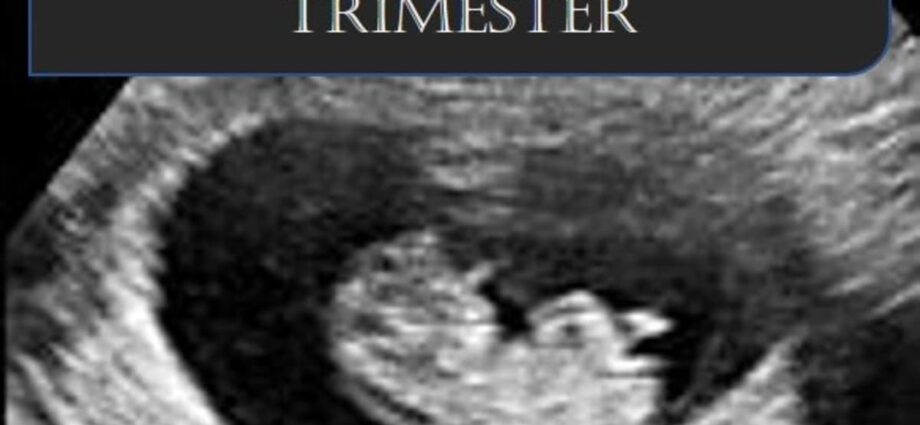
At the 4th week of pregnancy, the embryo is still very small.
At home, you can use the test. This is best done in the morning. After waking up, the concentration of hCG in the body is maximum. The test will show the most reliable result.
What happens during this period?
In size, the embryo resembles a poppy seed. Its length is only 4 mm. Weight does not exceed 1 g. Outwardly, its shape is similar to a flat disc. 3 embryonic petals have already formed. In the future, they will develop, forming organs and tissues.
The outer layer is called the ectoderm. It will form the basis of the uneven system. It will form the eye lenses, tooth enamel, skin and hair. From the middle layer – the mesoderm – the muscular frame, skeleton, connective tissues, as well as the excretory, reproductive, circulatory systems develop. The last layer of endoderm is necessary for the functioning of the digestion and endocrine glands.
The main work is now done by the paternal genes. They protect the fetus at the genetic level. This is necessary for the successful formation of important organs:
- Umbilical cord;
- Intestinal tube;
- The nervous system;
- Respiratory organs;
- Urinary system.
The embryo already has gills, as well as the rudiments of the limbs, mouth, eyes and nose. There is also a heart in the first stage of formation. It looks like a hollow tube. Blood flows through it in a direct stream. It is not yet possible to listen to the contractions of the heart. This can be done only 5-6 weeks after conception using ultrasound. The pulse is at least 100 beats per minute. Normally, the heart of the embryo beats at a frequency of 130 beats per minute.
Many changes occur with the embryo, which affects its structure.
The heart develops daily. Its tissues thicken, 2 chambers and a septum appear. The brain is forming at a rapid rate. It takes up about half of the neural tube. The rudiments of the hypothalamus are found in it. The spinal cord forms nerve nodes.
Changes in mother’s feelings
The first sign of pregnancy is delayed menstruation. The rest of the feelings are subjective.
If a woman’s nervous system is sensitive, she suffers from mood swings. Increased anxiety and irritability appear. Emotional uplift gives way to tearfulness. Due to the active development of the embryo, the stomach can pull. The pregnant woman is weak. Uterine discomfort makes it difficult to sit comfortably.
The breast responds to changes in the hormonal background. Its size increases slightly. Touching is unpleasant or painful. The nipple halos become darker and rougher.
Early toxicosis is very rare
Brown discharge is normal. This condition is called implantation bleeding. It arises from the introduction into the epithelial layer of the uterus of the embryo. Prolonged, growing heavy bleeding signals complications. You should consult a doctor.
Under the influence of progesterone, the production of vaginal secretions increases. It acquires a viscous and viscous structure. This is due to the formation of a mucous plug in the cervical canal, which will become a protective barrier for the fetus.
At such an early stage, ultrasound diagnostics can only be performed on a doctor’s prescription. It is performed with a transvaginal transducer. A small device is gently inserted into the vagina. This allows you to establish the place of attachment of the embryo. It looks like a tiny black spot on the scanner.
The study shows an increase in the corpus luteum. While a full-fledged placenta develops, the embryo feeds with it. It promotes the production of progesterone.
It takes a little time from conception to implantation.
A duplex scan will show dilation of the uterine vessels. This condition occurs due to the active nutrition of the embryo. Single veins can be observed around the endometrium, as well as changes in arterial blood flow.
Color Doppler ultrasonography will help identify pathologies and complications in the development of pregnancy. So at the initial stage, you can detect an ectopic and undeveloped pregnancy. The specialist will be able to rule out ovarian torsion or cystic drift. The attending physician decides on the need for such a study.
At this time, signs of pregnancy are weak. Until the time of the delay in menstruation, a woman is often unaware of her situation.